UK first for children with scoliosis
Posted on Friday 24th June 2016
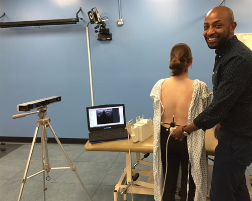
Clinical scientist, Eskinder Solomon, doing a scan at the gait laboratroy
Evelina London Children’s Hospital is developing the UK’s first 3D ultrasound imaging system for children with scoliosis that is safer than conventional techniques.
Scoliosis is a common medical condition in which a person’s spine is abnormally twisted and curved. Children and young people with severe scoliosis require X-rays every four to six months so that changes in their spine’s curvature can be monitored and surgery planned, if necessary.
The new imaging system pioneered at Evelina London’s ‘One Small Step' gait laboratory uses motion capture technology and ultrasound to generate 3D images of the spine’s shape. This is expected to help clinicians make better treatment decisions as they can see the spine in greater detail.
Eskinder Solomon, clinical scientist at Evelina London’s gait laboratory, says: “Our new imaging technique will provide clinicians with great detail on how a child’s scoliosis is progressing. We’re very pleased to be at the forefront of developing this technique at Evelina London.”
Daisy Biglands, 13, from Dorking in Surrey, has scoliosis and was one of the first young scoliosis patients to receive a scan using the new technology.
Daisy, who wears a back brace to help with her condition, says: “When I was diagnosed with scoliosis I was really scared. It’s not a nice thing to be told.
“The ultrasound was easy and took almost no time at all. Eskinder explained what was going to happen and even made it sound fun. The gel was cold and gooey but the scan was exciting. We could watch the 3D image of my spine build up as he scanned my back and I could see the curves really clearly.”
Scoliosis can develop at any age but is most common in children aged 10-15. Around three or four in every 1,000 children in the UK need treatment for scoliosis.
Jonathan Lucas, Daisy’s consultant spinal surgeon at Evelina London, says: “Children and young people with scoliosis require multiple X-rays of their spine as they grow so that we can monitor how the curve is progressing and appropriate treatment for patients like Daisy can be undertaken at the best possible time.
“Although the radiation doses are very low, it would best if they could be avoided. Unlike X-rays, 3D ultrasound scans are free of radiation. This pioneering technique is still in its infancy but may offer an even safer means of monitoring scoliosis.”