'Phantom' foot improves baby X-rays
Posted on Monday 2nd June 2014
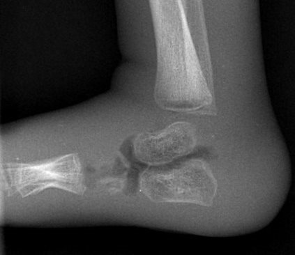
X-ray of the 'phantom' baby foot
Adam Jones, a clinical scientist in radiation safety, has worked out how to get better quality X-ray images of babies’ ankles, using existing equipment and without increasing the radiation dose.
Radiologists at Evelina London Children’s Hospital occasionally found that X-ray images of babies’ ankles were not good enough quality to help make decisions about their care. This is a common problem across the NHS.
“We couldn’t just increase the radiation dose to get a better image because UK regulations say patients’ exposure to radiation should be as low as possible whilst still getting a useable picture,” says Adam. “So first we had to check whether there was any other way we could improve the image quality.”
Adam tried setting up the equipment in different ways and took images of a ‘phantom’ baby’s foot, a model that trainee radiographers practice on.
The Evelina London radiologists and radiographers reviewed the results and agreed that one particular set up had significantly improved the image quality.
Adam believes this technique could also be used to improve X-ray image quality of other parts of the body such as hands, feet and elbows.
“Because this new process uses existing equipment it means we don’t have to invest anything extra and it’s quick to put into practice – we’ll soon be rolling it out across Evelina London for all X-rays of babies’ ankles,” says Adam.
Other NHS hospitals have already expressed an interest in using the technique after Adam presented his results at a national paediatric radiographer event hosted by Evelina London last November.
Adam won the ‘UK Young Professional Award’ at the recent Society for Radiological Protection international conference for this work. He next plans to investigate whether it is possible to keep the needed image quality while reducing the X-ray.